Microscope
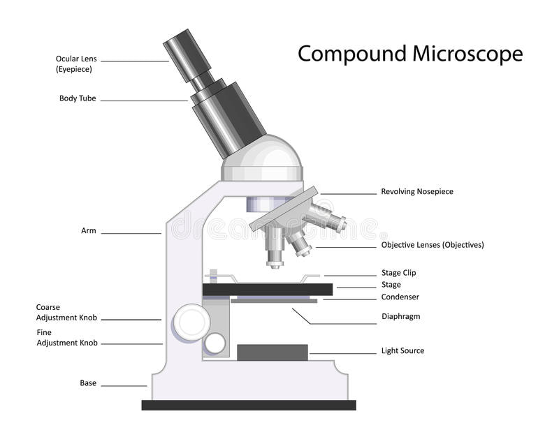
The microscope is used to enable us to see objects that are two small to be seen with the naked eye. In class we use the compound light microscope. A dissecting microscope can be used for observing living or dead organisms and they can be approximately as large as a fist. An electron microscope is the highest magnification microscope that we currently have. We cannot view live specimens under the electron microscope. To find the total magnification of the specimen under the microscope you must multiply the magnification of the ocular lens (eyepiece) and the objective lens (scanning, low, high power). Our microscopes in class have an ocular lens at 10x, a scanning objective at 4x, a low power objective at 10x and a high power objective at 40x. The respective total magnifications on scanning, low and high are 40x, 100x and 400x. Objects viewed under the microscope appear upside down and backwards. We never use the course adjustment under high power, it could break the slide or damage the lens. The diaphragm controls the amount of light on the slide. The field of view is largest and brightest under low power. Always carry the microscope using the arm and the base. When making a wet mount slide we place the cover slip at a 45 degree angle and slowly lower it down so that there are no air bubbles present in the slide.
The microscope is used to enable us to see objects that are two small to be seen with the naked eye. In class we use the compound light microscope. A dissecting microscope can be used for observing living or dead organisms and they can be approximately as large as a fist. An electron microscope is the highest magnification microscope that we currently have. We cannot view live specimens under the electron microscope. To find the total magnification of the specimen under the microscope you must multiply the magnification of the ocular lens (eyepiece) and the objective lens (scanning, low, high power). Our microscopes in class have an ocular lens at 10x, a scanning objective at 4x, a low power objective at 10x and a high power objective at 40x. The respective total magnifications on scanning, low and high are 40x, 100x and 400x. Objects viewed under the microscope appear upside down and backwards. We never use the course adjustment under high power, it could break the slide or damage the lens. The diaphragm controls the amount of light on the slide. The field of view is largest and brightest under low power. Always carry the microscope using the arm and the base. When making a wet mount slide we place the cover slip at a 45 degree angle and slowly lower it down so that there are no air bubbles present in the slide.

Gel Electrophoresis
Gel Electrophoresis uses an electircal current to separate fragments of DNA. Used in criminal investigations, paternity tests and evoluntionary relationships.
To create fragments of DNA, the DNA is mixed with special cutting substances called restrictive enzymes. It is also mixed with a special dye so that it can be seen on the gel. The mixture is then loaded into wells on an agarose gel. An electric current is then applied and fragments move through the gel. The smaller pieces move further and the larger pieces don’t move as far. Results show a banding pattern on the gel under special light.


Chromatography
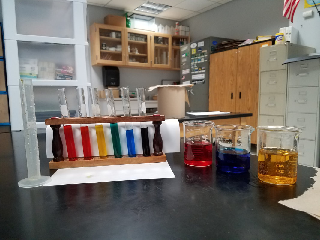
Chromatography is a technique used to separate a mixture of molecules, generally different pigments. The set up includes a beaker with a solvent and special chromatography paper for the separation. A common example of this technique would involve putting a concentrated amount of chlorophyll (a green coloring found in plants) extract on a piece of filter paper and then placing the filter paper with the extract into a solvent.As the solvent soaks up into the paper and moves upward, colors that exist within the chlorophyll are separated into bands of color on the filter paper.
Chromatography is a technique used to separate a mixture of molecules, generally different pigments. The set up includes a beaker with a solvent and special chromatography paper for the separation. A common example of this technique would involve putting a concentrated amount of chlorophyll (a green coloring found in plants) extract on a piece of filter paper and then placing the filter paper with the extract into a solvent.As the solvent soaks up into the paper and moves upward, colors that exist within the chlorophyll are separated into bands of color on the filter paper.
 |  |
 Centrifuge A centrifuge separates mixtures such as blood components or cell parts based on density |  |

Tissue Culturing
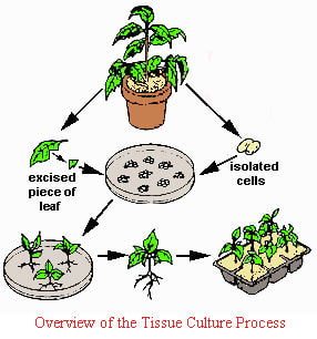
Tissue culturing is a technique used in growing living tissue and microorganisms such as bacteria in the lab. Some examples include in vitro fertilization, bacterial culture (strep throat) and even cloning.
What is tissue culturing good for?
Tissue culturing is a technique used in growing living tissue and microorganisms such as bacteria in the lab. Some examples include in vitro fertilization, bacterial culture (strep throat) and even cloning.
What is tissue culturing good for?
- Can be used to grow either animal or plant tissue
- Can grow a large amount of certain types of organisms
- Can be used to produce large numbers of the same cells or the same organisms (cloning)
- Can also be used to grow tissue for analysis (ex.- cancer)
- Can even be used to grow whole entire organs!
Staining
This technique makes certain cell parts visible depending on the stain being used. Staining kills the specimen!!!!
Indicators
Indicators are substances that change color when they encounter specific chemical conditions
Examples:
This technique makes certain cell parts visible depending on the stain being used. Staining kills the specimen!!!!
Indicators
Indicators are substances that change color when they encounter specific chemical conditions
Examples:
- Lugol’s Iodine is an indicator of starch. It goes from amber to purple/black when in the presence of a starch
- Benedict’s solution is an indicator for glucose. It starts a light blue color and changes to brick orange when mixed with glucose. MUST be heated for the change to occur
- pH indicators (litmus paper, strips or fluids) show a color change based on how acidic or basic a substance is. The color change should be compared to a known color scale from the package of the pH indicator.
Classification
We use this system called classification to group together living things with similar characteristics. Groups are arranged from most general to most specific, the more general the group the greater the number of organisms that belong. Classification is based on structures.
Taxonomy
The science of classifying and naming organisms. This system that was developed by a Swedish scientist, Carolus Linnaeus, is binomial nomenclature. It uses 2 names (the Genus and the species name) to make up the scientific name. Genus is always capitalized and species is always lower case. When written the scientific name is always Italicized or underlined. For example: The scientific name for a dog is Canis familiaris and the scientific name for humans is Homo sapiens.
Dichotomous key
Used to classify organisms that are similar. Always between two options
Used to classify organisms that are similar. Always between two options